Why Choose Orthopedic Surgical Plates for Better Outcomes?

Why Choose Orthopedic Surgical Plates for Better Outcomes?

Fractures remain a major global health burden. The 2021 Global Burden of Disease study, published in The Lancet, recorded about 178 million new fractures worldwide in 2019. It also estimated 455 million people living with fracture-related conditions. These figures show why reliable fixation matters in operating rooms.

Orthopedic surgical plates can support alignment, stability, and earlier functional recovery when selected correctly. Their value depends on bone quality, fracture pattern, soft-tissue condition, and surgical technique. A plate is not a complete solution. Professor Stephan Perren, a leading AO Foundation researcher, emphasized this principle: “The implant is a tool, not the treatment.”

That distinction matters.

Recent analyses from Grand View Research and MarketsandMarkets identify trauma fixation as a continuing growth area within the orthopedic devices industry. However, commercial forecasts use different definitions and should not replace clinical evidence. The American Academy of Orthopaedic Surgeons stresses individualized assessment, careful planning, and appropriate follow-up.

In practice, better outcomes may involve a contoured plate sitting beneath thin tissue, stable screws holding a difficult fragment, and rehabilitation beginning at the right moment. Small choices can change recovery.

Yet plates can fail.

Poor implant selection, infection, nonunion, or excessive stiffness may compromise results. Surgeons must balance mechanical stability with biological preservation. Patients also need clear explanations about risks, recovery time, and realistic expectations. This article examines how orthopedic surgical plates work, where they help most, and why thoughtful clinical judgment remains essential.

Why Choose Orthopedic Surgical Plates for Better Outcomes?

What Are Orthopedic Surgical Plates and How Do They Work?

Why Choose Orthopedic Surgical Plates for Better Outcomes?

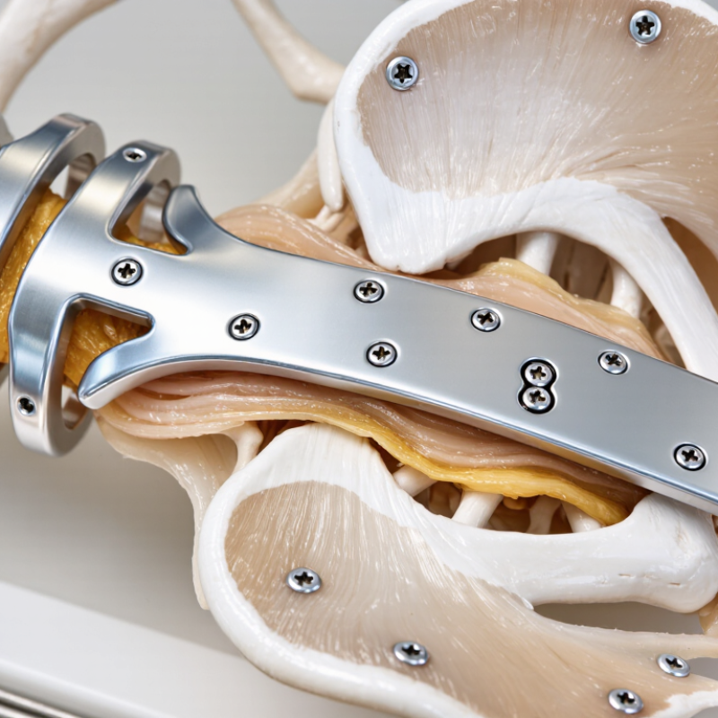
Orthopedic surgical plates are shaped metal implants placed beside a fractured bone. They hold broken segments in a controlled position while healing occurs. Most plates work with screws, which connect the plate and bone like a temporary external support. Some designs compress the fracture line. Others preserve small gaps and provide relative stability for new bone formation.

The need is substantial. The Global Burden of Disease Study 2019 reported about 178 million new fractures worldwide that year. It also estimated 455 million people living with fracture-related symptoms. These figures, published in the Journal of Bone and Mineral Research, show why dependable fixation matters. In practice, surgeons assess bone quality, fracture pattern, soft-tissue condition, and movement demands before selecting a plate. A plate is not a cure. Poor positioning, infection, delayed healing, or excessive loading can still affect recovery. Evidence is not uniform for every fracture type.

Tips: Ask how the plate will support alignment and movement. Clarify weight-bearing limits. Follow wound-care instructions closely. Report increasing warmth, drainage, numbness, or unusual pain promptly. Do not treat online recovery timelines as personal medical advice. Even small details, such as swelling after a long walk, deserve attention.

How Plate Design Supports Bone Stability and Healing

Orthopedic surgical plates help stabilize fractured bones while the body builds new tissue. Their value depends heavily on design, not metal strength alone. A well-contoured plate follows the bone’s surface, reducing gaps and unnecessary pressure on nearby soft tissue. That fit can support alignment during movement and routine rehabilitation. Small details matter.

Locking holes allow screws to connect firmly with the plate, creating a stable fixed-angle construct. This can help in fragile bone or fractures with limited surface contact. Compression holes may draw bone fragments together when direct healing is clinically appropriate. Some designs reduce contact beneath the plate, helping preserve the local biological environment. Surgeons still choose screw position, length, and sequence according to fracture pattern and bone quality. A plate cannot correct poor reduction.

In practice, imaging and hands-on assessment guide whether the construct is too flexible or excessively stiff. Too much motion may delay healing, while excessive rigidity can shift stress away from the bone. The best design balances stability, controlled movement, and respect for surrounding tissue. It also supports practical needs, including clear imaging and manageable placement through a limited incision. No design works equally well for every patient. Age, smoking, infection risk, activity, and fracture location can change the plan. Some outcomes remain unpredictable, and follow-up matters.

When Orthopedic Surgeons Choose Plates for Fracture Treatment

Why Choose Orthopedic Surgical Plates for Better Outcomes?

When orthopedic surgeons choose plates for fracture treatment, they assess more than the broken bone. They study the fracture pattern, bone quality, soft-tissue condition, and the patient’s daily needs. A plate may support alignment when the fracture is unstable or crosses a joint. It can also help restore length, rotation, and function.

The decision is not always clear. Plain X-rays may hide small fragments or joint involvement, so surgeons may use additional imaging. During surgery, they check reduction, plate position, screw direction, and soft-tissue protection. Stable fixation can support earlier movement, especially around the wrist, ankle, or elbow. Early motion matters.

Plate selection requires judgment.

Clinical experience guides this process, but experience alone is not enough. Surgeons also consider published evidence, patient risks, and the limits of each fixation method. Poor bone quality, infection, excessive implant prominence, or delayed healing can affect results. A plate that looks ideal on an X-ray may still irritate nearby tissue. That detail is easy to underestimate. Careful planning, accurate placement, follow-up imaging, and rehabilitation help reduce preventable problems. Still, healing varies, and no surgical plan guarantees a perfect outcome.

Which Materials and Features Improve Surgical Outcomes?

Orthopedic surgical plates perform best when their material matches the fracture and the patient’s biology. Titanium offers a lower elastic modulus than stainless steel, which may reduce stress shielding around healing bone. It is also lighter and highly corrosion resistant. Stainless steel remains strong, affordable, and familiar in trauma settings. However, material choice alone cannot guarantee better healing. A 2022 Cochrane review on distal-radius fracture surgery reported low- to moderate-certainty evidence, with no consistent long-term functional advantage for one fixation method. That finding deserves attention.

Locking screws can improve stability in weak, osteoporotic bone. Variable-angle holes help surgeons avoid sensitive joint surfaces and adapt screw direction. A low-profile plate may reduce tendon irritation, especially near the wrist or ankle. Rounded edges and anatomical contouring also matter. Yet excessive rigidity can create stress concentration, while poor contouring may irritate soft tissue. No plate is perfect. Surgical experience, reduction quality, blood supply, and rehabilitation often influence outcomes more than a feature list. The American Academy of Orthopaedic Surgeons continues to emphasize patient-specific planning and shared clinical judgment in fracture care guidance.

Tips: Ask whether titanium or stainless steel suits the fracture pattern. Check the plate’s locking options and screw-angle range. Confirm imaging shows joint clearance. Review evidence, not marketing language. Even experienced teams should question whether a thinner plate truly improves comfort without reducing stability.

Why Choose Orthopedic Surgical Plates for Better Outcomes? — Which Materials and Features Improve Surgical Outcomes?

Material or Design Dimension Typical Options Relevant Characteristics How It May Support Surgical Outcomes Important Clinical Considerations
Implant Material Titanium alloy
Commonly used for internal fixation
High strength-to-weight ratio, good corrosion resistance, and a modulus of elasticity generally closer to cortical bone than stainless steel. Can provide stable fixation while potentially reducing stress concentration at the bone–implant interface. Its radiographic characteristics may also make fracture assessment easier than with more radiopaque metals. Material selection should consider fracture pattern, load requirements, bone quality, anatomical site, and surgeon experience. Titanium is not automatically superior for every indication.
Implant Material Stainless steel
Commonly used in trauma fixation
High strength, good fatigue resistance, established manufacturing history, and reliable performance in many fixation applications. Its mechanical strength can support fixation in areas exposed to substantial loading when the plate is appropriately selected and applied. It is generally stiffer and more radiopaque than titanium. Compatibility with other implanted metals should be assessed to limit concerns related to galvanic corrosion.
Implant Material Cobalt–chromium alloy
Selected high-strength applications
High strength, high hardness, and strong wear and corrosion resistance. May be considered when high mechanical strength and resistance to deformation are important. Its relatively high stiffness and hardness can complicate contouring and may increase stress shielding concerns in selected situations. Application-specific evidence is required.
Plate Geometry Anatomically pre-contoured plate Designed to follow the typical shape of a particular bone region, reducing the amount of intraoperative bending required. May shorten contouring time, help preserve plate strength, and improve fit when the patient’s anatomy matches the intended design. Pre-contouring does not replace fracture reduction or individual anatomical assessment. Excessive intraoperative bending can weaken some plate designs.
Fixation Principle Locking screw compatibility Locking screws engage the plate, creating a fixed-angle construct that does not depend entirely on plate-to-bone compression. Can be useful in osteoporotic bone, metaphyseal fractures, and situations where preservation of periosteal blood supply is desirable. Locking technology does not eliminate the need for accurate reduction. Overly rigid constructs or inappropriate screw distribution may affect healing.
Fixation Principle Combination holes Plate holes can accommodate conventional compression screws, locking screws, or both, depending on the design. Provides flexibility to combine compression, bridging, and fixed-angle fixation techniques within one plate. The selected screw type, sequence, and position should match the desired biomechanical function of each part of the construct.
Soft-Tissue Consideration Low-profile design Reduces implant prominence while maintaining the required mechanical function. May reduce irritation from soft-tissue prominence, particularly in areas with limited tissue coverage. A thinner plate is not necessarily appropriate for a high-load fracture. Mechanical requirements must be balanced with soft-tissue considerations.
Bone–Plate Interface Limited-contact or undercut surface Minimizes the area of direct contact between the plate and the outer bone surface compared with traditional broad-contact designs. May help preserve local cortical blood supply and reduce unnecessary disruption of the periosteal interface. Clinical benefit depends on surgical technique, reduction quality, soft-tissue handling, and the overall stability of the construct.
Mechanical Performance Fatigue-resistant design Plate thickness, cross-sectional geometry, material properties, screw-hole configuration, and surface finish influence resistance to cyclic loading. Appropriate fatigue strength can reduce the risk of plate deformation or fatigue failure while the fracture heals. Laboratory fatigue performance cannot predict every clinical result. Nonunion, excessive loading, poor reduction, and improper screw placement can increase failure risk.
Screw Options Multiple screw diameters and lengths A range of screw sizes allows fixation to be adapted to bone thickness, fracture location, and available purchase. Supports more appropriate screw selection and may reduce the risk of inadequate fixation or penetration of nearby joints and soft tissues. Screw length and trajectory should be confirmed with appropriate imaging and anatomical landmarks. More screws do not always produce a better construct.
Imaging and Navigation Radiographic visualization features Plate shape, radiopacity, and dedicated reference holes can assist intraoperative imaging and postoperative assessment. Clear visualization can support confirmation of alignment, screw position, joint clearance, and fracture healing. Imaging quality depends on positioning, equipment, fracture location, and surgical technique. It should be combined with clinical evaluation.
Biocompatibility Implant-grade, corrosion-resistant material Materials intended for implantation must meet applicable medical-device requirements for biocompatibility, manufacturing quality, and mechanical performance. Reduces the likelihood of material-related complications when the implant is used as intended and the patient is appropriately assessed. Patient history, known metal sensitivity, infection risk, systemic health, and concurrent implants should be considered before surgery.
Clinical Fit Anatomy- and fracture-specific selection Plate choice should reflect the bone involved, fracture morphology, bone quality, soft-tissue condition, expected loading, and treatment objectives. Matching the plate and fixation strategy to the clinical problem is more important than selecting a material or feature in isolation. Outcomes are also influenced by reduction, soft-tissue preservation, infection prevention, rehabilitation, patient compliance, and follow-up care.

Note: The information above summarizes general orthopedic fixation principles. Final implant and surgical-technique decisions should be made by a qualified orthopedic surgeon according to the patient’s anatomy, fracture pattern, bone quality, and applicable clinical guidance.

What Factors Affect Recovery After Plate Fixation?

Why Choose Orthopedic Surgical Plates for Better Outcomes?

Recovery after plate fixation depends on more than the plate itself. The fracture’s location, pattern, bone quality, and soft-tissue damage can influence healing time. A stable plate may support alignment, but it cannot replace healthy biology. Blood supply around the fracture remains important.

Pain matters. In the first weeks, swelling may increase after activity, especially around the ankle, wrist, or elbow. Elevating the limb and following the surgeon’s weight-bearing instructions can reduce stress on healing tissues.

Physical therapy often begins with gentle movement, then gradually adds strength and balance exercises. Moving too little may cause stiffness. Moving too aggressively may irritate the incision or fracture.

Smoking and nicotine can reduce blood flow and delay bone healing. Diabetes, poor nutrition, infection, and some medications may also affect recovery. Adequate protein, calcium, vitamin D, sleep, and hydration support the body’s repair process, but supplements should follow professional advice. Follow-up X-rays help clinicians judge alignment and callus formation. A normal-looking wound does not always prove complete healing.

Recovery is rarely linear. Progress can stall. Patients should report increasing redness, drainage, fever, numbness, severe pain, or a suddenly loose feeling around the fixation. These signs need prompt medical assessment. Return to work, driving, sports, or heavy lifting should depend on clinical examination rather than confidence alone. Even careful patients may heal slowly, and recovery plans sometimes need revision.